Non-invasive ultrasound imaging to evaluate the heart’s structure and function in real time.
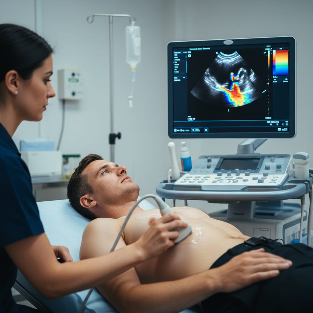
Echocardiography (ECHO) uses high-frequency sound waves (ultrasound) to create moving images of the heart. It helps physicians assess the size, shape, structure, and pumping action of the heart valves and chambers, detecting conditions that might be missed in a standard checkup.
A simple 3-step process designed for your convenience.
A trained technician applies conductive gel and places probe on your chest.
Ultrasound echoes are converted into live images showing heart chambers.
A cardiologist reviews images and Doppler data to guide your treatment.
Completely painless test without radiation, using only sound waves.
Instant visualization of valve function and chamber motion.
Detects structural heart disease, fluid around heart, and more.
Essential for pre/post-operative assessment and long-term care management.